Abstract
Clinical Image
Pulmonary Infarction Mimicking An Aspergilloma In A Heart Transplant Recipient
Antonacci F*, Belliato M, Bortolotto C, Di Perna D, Dore R, Orlandoni G and D’Armini AM
Published: 30 January, 2017 | Volume 1 - Issue 1 | Pages: 005-006
his patient (male, 59 years old) underwent cardiac re-transplantation for chronic rejection. Prior to re-transplantation, the patient was in NYHA class IV, with a clear chest x ray. On 14th postoperative day, he presented hemoptysis. On chest x-ray, a left lower lobe opacity was seen. Therefore, a chest CT scan was done and it showed a round mass within a pulmonary cavity surrounded by airspace in proximity of the pulmonary artery.
Read Full Article HTML DOI: 10.29328/journal.jcmei.1001002 Cite this Article Read Full Article PDF
Keywords:
Pulmonary aspergillosis; Lobectomy; Pulmonary infarction
References
- Franquet T, Müller NL, Giménez A, Guembe P, de La Torre J, et al. Spectrum of pulmonary aspergillosis: histologic, clinical, and radiologic findings. Radiographics. 2001; 21: 825-837. Ref.: https://goo.gl/yIgqMZ
- Bray TJ, Mortensen KH, Gopalan D. Multimodality imaging of pulmonary infarction. Eur J Radiol. 2014; 83: 2240-2254. Ref.: https://goo.gl/R8RTCr
Figures:
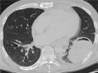
Figure 1
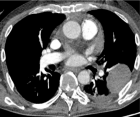
Figure 2
Similar Articles
Recently Viewed
-
A Comparative Analysis of Traditional Latent Fingerprint Visualization Methods and Innovative Silica Gel G Powder ApproachBhoomi Aggarwal*. A Comparative Analysis of Traditional Latent Fingerprint Visualization Methods and Innovative Silica Gel G Powder Approach. J Forensic Sci Res. 2024: doi: 10.29328/journal.jfsr.1001063; 8: 040-046
-
A Comparative Study of Metoprolol and Amlodipine on Mortality, Disability and Complication in Acute StrokeJayantee Kalita*,Dhiraj Kumar,Nagendra B Gutti,Sandeep K Gupta,Anadi Mishra,Vivek Singh. A Comparative Study of Metoprolol and Amlodipine on Mortality, Disability and Complication in Acute Stroke. J Neurosci Neurol Disord. 2025: doi: 10.29328/journal.jnnd.1001108; 9: 039-045
-
Huge Mucinous Cyst Adenoma, Case Report SeriesAli Mohammed Ali Elimam*,Isam Mohamed Babbiker,Reem Mohammed Elhaj Farah,Ayat Salih Abbas,Omer Mohamed Abubeker Sayed. Huge Mucinous Cyst Adenoma, Case Report Series. Clin J Obstet Gynecol. 2025: doi: 10.29328/journal.cjog.1001184; 8: 019-022
-
Does change in cervical dilation after anesthesia impact latency after cerclage placement?Michelle N Lende*, Paul J Feustel, Erica K Nicasio, Tara A Lynch. Does change in cervical dilation after anesthesia impact latency after cerclage placement?. Clin J Obstet Gynecol. 2023: doi: 10.29328/journal.cjog.1001125; 6: 028-031
-
Minimally invasive cytoreductive surgery in advanced ovarian cancer: A nonselected consecutive series of robotic-assisted casesNatalie Shammas*, Rosa Avila, Christopher Khatchadourian, Erland Laurence Spencer-Smith, Lisa Stern, Steven Vasilev. Minimally invasive cytoreductive surgery in advanced ovarian cancer: A nonselected consecutive series of robotic-assisted cases. Clin J Obstet Gynecol. 2023: doi: 10.29328/journal.cjog.1001126; 6: 032-037
Most Viewed
-
Evaluation of Biostimulants Based on Recovered Protein Hydrolysates from Animal By-products as Plant Growth EnhancersH Pérez-Aguilar*, M Lacruz-Asaro, F Arán-Ais. Evaluation of Biostimulants Based on Recovered Protein Hydrolysates from Animal By-products as Plant Growth Enhancers. J Plant Sci Phytopathol. 2023 doi: 10.29328/journal.jpsp.1001104; 7: 042-047
-
Sinonasal Myxoma Extending into the Orbit in a 4-Year Old: A Case PresentationJulian A Purrinos*, Ramzi Younis. Sinonasal Myxoma Extending into the Orbit in a 4-Year Old: A Case Presentation. Arch Case Rep. 2024 doi: 10.29328/journal.acr.1001099; 8: 075-077
-
Feasibility study of magnetic sensing for detecting single-neuron action potentialsDenis Tonini,Kai Wu,Renata Saha,Jian-Ping Wang*. Feasibility study of magnetic sensing for detecting single-neuron action potentials. Ann Biomed Sci Eng. 2022 doi: 10.29328/journal.abse.1001018; 6: 019-029
-
Pediatric Dysgerminoma: Unveiling a Rare Ovarian TumorFaten Limaiem*, Khalil Saffar, Ahmed Halouani. Pediatric Dysgerminoma: Unveiling a Rare Ovarian Tumor. Arch Case Rep. 2024 doi: 10.29328/journal.acr.1001087; 8: 010-013
-
Physical activity can change the physiological and psychological circumstances during COVID-19 pandemic: A narrative reviewKhashayar Maroufi*. Physical activity can change the physiological and psychological circumstances during COVID-19 pandemic: A narrative review. J Sports Med Ther. 2021 doi: 10.29328/journal.jsmt.1001051; 6: 001-007

HSPI: We're glad you're here. Please click "create a new Query" if you are a new visitor to our website and need further information from us.
If you are already a member of our network and need to keep track of any developments regarding a question you have already submitted, click "take me to my Query."


