Abstract
Clinical Image
The Death of a Baby from the Congenital Anomalies of the Urinary Tract
Astrit Gashi M*, Gent Sopa, Ilir Kadiri, Majlinda Bala and Petrit Pupa
Published: 22 February, 2018 | Volume 2 - Issue 1 | Pages: 001-002
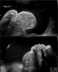
A 36-year-old woman pregnant, G2 P1, presented at 27 weeks of gestation after two previous visits elsewhere, as an outpatient in a gynecological clinic. An ultrasound examination revealed bilateral hydronephrosis. Also, ureteral dilation and bladder overdistension was present (Figures 1-3). We evaluated that the cause was a urinary tract obstruction. Specifically, we are dealing with posterior urethral valves. The anteroposterior diameter of the pelvis on a transverse view of the abdomen was 6 mm. The amniotic fluid index (AFI) was 3 cm, so, oligohydramnios.
Read Full Article HTML DOI: 10.29328/journal.jcmei.1001009 Cite this Article Read Full Article PDF
References
Figures:
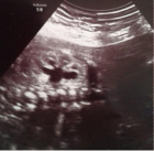
Figure 1
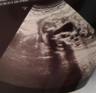
Figure 2
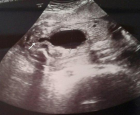
Figure 3
Similar Articles
-
Cystic Micronodular Thymoma. Report of a CaseMona Mlika*,Adel Marghli ,Faouzi Mezni. Cystic Micronodular Thymoma. Report of a Case . . 2017 doi: 10.29328/journal.jcmei.1001001; 1: 001-004
-
Pulmonary Infarction Mimicking An Aspergilloma In A Heart Transplant RecipientAntonacci F*,Belliato M,Bortolotto C,Di Perna D,Dore R,Orlandoni G,D’Armini AM. Pulmonary Infarction Mimicking An Aspergilloma In A Heart Transplant Recipient . . 2017 doi: 10.29328/journal.jcmei.1001002; 1: 005-006
-
Recurrent Peripheral Ameloblastoma of the Mandible: A Case ReportAngela Jordão Camargo*,Mayara Cheade,Celso Martinelli,Plauto Christopher Aranha Watanabe. Recurrent Peripheral Ameloblastoma of the Mandible: A Case Report. . 2017 doi: 10.29328/journal.jcmei.1001003; 1: 007-010
-
Tumours of the Uterine Corpus: A Histopathological and Prognostic Evaluation Preliminary of 429 PatientsJorge F Cameselle-Teijeiro*,Javier Valdés-Pons,Lucía Cameselle-Cortizo,Isaura Fernández-Pérez,MaríaJosé Lamas-González,Sabela Iglesias-Faustino,Elena Figueiredo Alonso,María-Emilia Cortizo-Torres,María-Concepción Agras-Suárez,Araceli Iglesias-Salgado,Marta Salgado-Costas,Susana Friande-Pereira,Fernando C Schmitt. Tumours of the Uterine Corpus: A Histopathological and Prognostic Evaluation Preliminary of 429 Patients . . 2017 doi: 10.29328/journal.jcmei.1001004; 1: 011-019
-
The Risk Factors for Ankle Sprain in Cadets at a Male Military School in Iran: A Retrospective Case-control StudyFarzad Najafipour*,Farshad Najafipour,Mohammad Hassan Majlesi,Milad Darejeh. The Risk Factors for Ankle Sprain in Cadets at a Male Military School in Iran: A Retrospective Case-control Study. . 2017 doi: 10.29328/journal.jcmei.1001005; 1: 020-026
-
Magnetic Resonance Imaging Can Detect Symptomatic Patients with Facet Joint Pain. A Retrospective AnalysisWolfgang Freund*,Frank Weber,Reinhard Meier,Stephan Klessinger. Magnetic Resonance Imaging Can Detect Symptomatic Patients with Facet Joint Pain. A Retrospective Analysis. . 2017 doi: 10.29328/journal.jcmei.1001006; 1: 027-036
-
Secondary Onychomycosis Development after Cosmetic Procedure-Case ReportMariusz Dyląg*,Emilia Flisowska,Patryk Bielecki,Maria Kozioł-Gałczyńska,Weronika Jasińska. Secondary Onychomycosis Development after Cosmetic Procedure-Case Report . . 2017 doi: 10.29328/journal.jcmei.1001007; 1: 037-045
-
Andy Gump deformityPirabu Sakthivel*,Chirom Amit Singh,Chandra Sharma. Andy Gump deformity. . 2017 doi: 10.29328/journal.jcmei.1001008; 1: 046-047
-
The Death of a Baby from the Congenital Anomalies of the Urinary TractAstrit Gashi M*,Gent Sopa,Ilir Kadiri,Majlinda Bala,Petrit Pupa. The Death of a Baby from the Congenital Anomalies of the Urinary Tract. . 2018 doi: 10.29328/journal.jcmei.1001009; 2: 001-002
-
New technique of imaging cellular change to squmous cells metaplsia of cervixSalwa Samir Anter*. New technique of imaging cellular change to squmous cells metaplsia of cervix . . 2019 doi: 10.29328/journal.jcmei.1001010; 3: 001-001
Recently Viewed
-
Physiotherapy Undergraduate Students’ Perception About Clinical Education; A Qualitative StudyPravakar Timalsina*,Bimika Khadgi. Physiotherapy Undergraduate Students’ Perception About Clinical Education; A Qualitative Study. J Nov Physiother Rehabil. 2024: doi: 10.29328/journal.jnpr.1001063; 8: 043-052
-
Clinical Significance of Anterograde Angiography for Preoperative Evaluation in Patients with Varicose VeinsYi Liu,Dong Liu#,Junchen Li#,Tianqing Yao,Yincheng Ran,Ke Tian,Haonan Zhou,Lei Zhou,Zhumin Cao*,Kai Deng*. Clinical Significance of Anterograde Angiography for Preoperative Evaluation in Patients with Varicose Veins. J Radiol Oncol. 2025: doi: 10.29328/journal.jro.1001073; 9: 001-006
-
Regional Anesthesia Challenges in a Pregnant Patient with VACTERL Association: A Case ReportUzma Khanam*,Abid,Bhagyashri V Kumbar. Regional Anesthesia Challenges in a Pregnant Patient with VACTERL Association: A Case Report. Int J Clin Anesth Res. 2025: doi: 10.29328/journal.ijcar.1001027; 9: 010-012
-
Cystoid Macular Oedema Secondary to Bimatoprost in a Patient with Primary Open Angle GlaucomaKonstantinos Kyratzoglou*,Katie Morton. Cystoid Macular Oedema Secondary to Bimatoprost in a Patient with Primary Open Angle Glaucoma. Int J Clin Exp Ophthalmol. 2025: doi: 10.29328/journal.ijceo.1001059; 9: 001-003
-
Navigating Neurodegenerative Disorders: A Comprehensive Review of Current and Emerging Therapies for Neurodegenerative DisordersShashikant Kharat*, Sanjana Mali*, Gayatri Korade, Rakhi Gaykar. Navigating Neurodegenerative Disorders: A Comprehensive Review of Current and Emerging Therapies for Neurodegenerative Disorders. J Neurosci Neurol Disord. 2024: doi: 10.29328/journal.jnnd.1001095; 8: 033-046
Most Viewed
-
Evaluation of Biostimulants Based on Recovered Protein Hydrolysates from Animal By-products as Plant Growth EnhancersH Pérez-Aguilar*, M Lacruz-Asaro, F Arán-Ais. Evaluation of Biostimulants Based on Recovered Protein Hydrolysates from Animal By-products as Plant Growth Enhancers. J Plant Sci Phytopathol. 2023 doi: 10.29328/journal.jpsp.1001104; 7: 042-047
-
Sinonasal Myxoma Extending into the Orbit in a 4-Year Old: A Case PresentationJulian A Purrinos*, Ramzi Younis. Sinonasal Myxoma Extending into the Orbit in a 4-Year Old: A Case Presentation. Arch Case Rep. 2024 doi: 10.29328/journal.acr.1001099; 8: 075-077
-
Feasibility study of magnetic sensing for detecting single-neuron action potentialsDenis Tonini,Kai Wu,Renata Saha,Jian-Ping Wang*. Feasibility study of magnetic sensing for detecting single-neuron action potentials. Ann Biomed Sci Eng. 2022 doi: 10.29328/journal.abse.1001018; 6: 019-029
-
Pediatric Dysgerminoma: Unveiling a Rare Ovarian TumorFaten Limaiem*, Khalil Saffar, Ahmed Halouani. Pediatric Dysgerminoma: Unveiling a Rare Ovarian Tumor. Arch Case Rep. 2024 doi: 10.29328/journal.acr.1001087; 8: 010-013
-
Physical activity can change the physiological and psychological circumstances during COVID-19 pandemic: A narrative reviewKhashayar Maroufi*. Physical activity can change the physiological and psychological circumstances during COVID-19 pandemic: A narrative review. J Sports Med Ther. 2021 doi: 10.29328/journal.jsmt.1001051; 6: 001-007

HSPI: We're glad you're here. Please click "create a new Query" if you are a new visitor to our website and need further information from us.
If you are already a member of our network and need to keep track of any developments regarding a question you have already submitted, click "take me to my Query."


