Figure 1
Cystic Micronodular Thymoma. Report of a Case
Mona Mlika*, Adel Marghli and Faouzi Mezni
Published: 20 January, 2017 | Volume 1 - Issue 1 | Pages: 001-004
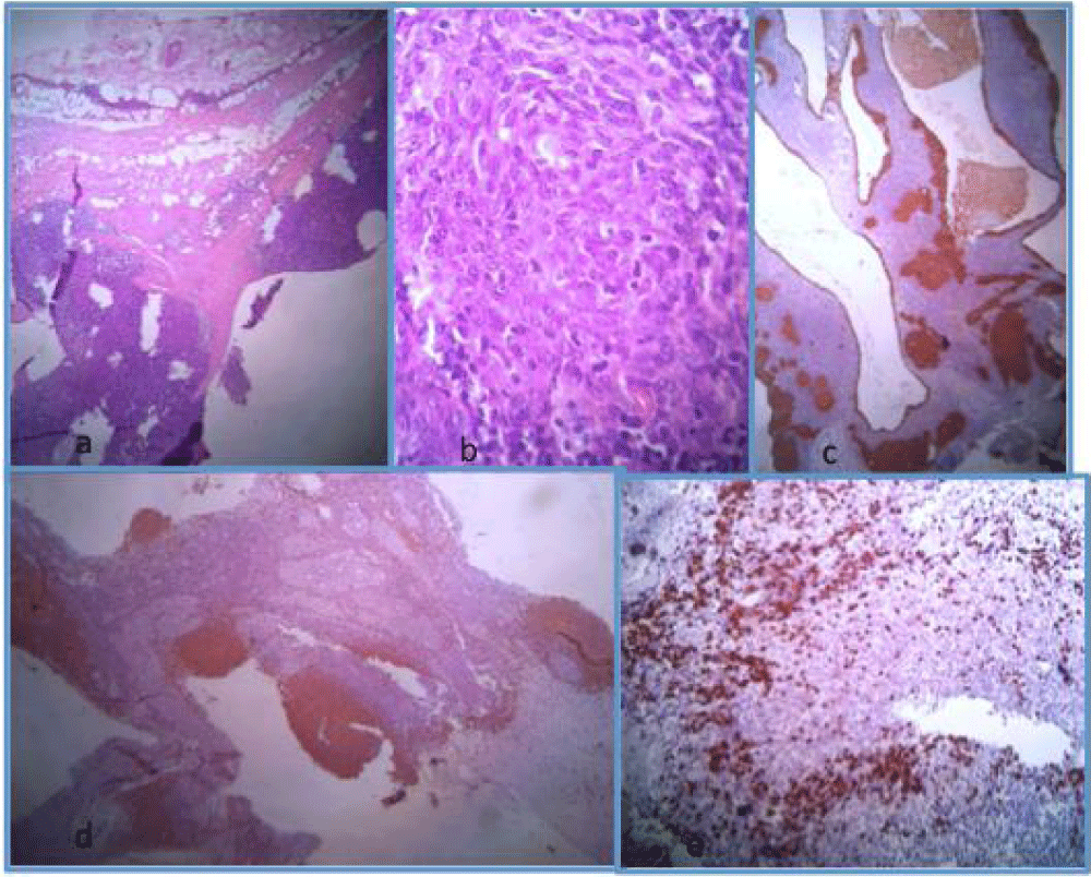
Figure 1:
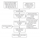
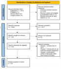
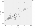
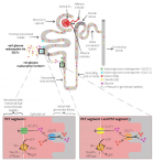
1: (a) Well limited tumour partially cystic made of epithelial nests and an abundant lymphoid stroma (HE x 250), (b) The epithelial cells are of spindle shape (HE x 400), (c) Immunohistochemical study using the cytokeratin antibody and highlighting the epithelial nests (HE x 250), (d) Immunohistochemical study using the CD20 antibody and highlighting the lymphoid follicles (HE x 250), (e) Immunohistochemical study using the Tdt antibody and highlighting the immature lymphocytes (HE x 250).
Read Full Article HTML DOI: 10.29328/journal.jcmei.1001001 Cite this Article Read Full Article PDF
More Images
Similar Articles
-
Cystic Micronodular Thymoma. Report of a CaseMona Mlika*,Adel Marghli ,Faouzi Mezni. Cystic Micronodular Thymoma. Report of a Case . . 2017 doi: 10.29328/journal.jcmei.1001001; 1: 001-004
-
Tumours of the Uterine Corpus: A Histopathological and Prognostic Evaluation Preliminary of 429 PatientsJorge F Cameselle-Teijeiro*,Javier Valdés-Pons,Lucía Cameselle-Cortizo,Isaura Fernández-Pérez,MaríaJosé Lamas-González,Sabela Iglesias-Faustino,Elena Figueiredo Alonso,María-Emilia Cortizo-Torres,María-Concepción Agras-Suárez,Araceli Iglesias-Salgado,Marta Salgado-Costas,Susana Friande-Pereira,Fernando C Schmitt. Tumours of the Uterine Corpus: A Histopathological and Prognostic Evaluation Preliminary of 429 Patients . . 2017 doi: 10.29328/journal.jcmei.1001004; 1: 011-019
-
Secondary Onychomycosis Development after Cosmetic Procedure-Case ReportMariusz Dyląg*,Emilia Flisowska,Patryk Bielecki,Maria Kozioł-Gałczyńska,Weronika Jasińska. Secondary Onychomycosis Development after Cosmetic Procedure-Case Report . . 2017 doi: 10.29328/journal.jcmei.1001007; 1: 037-045
-
Occipital lobe ependymal cyst with unusual presentationOkacha Naama*,Abdelilah Idir,Omar Boulahroud . Occipital lobe ependymal cyst with unusual presentation. . 2019 doi: 10.29328/journal.jcmei.1001012; 3: 009-011
-
CT-guided Retrograde Urography as a Diagnostic Tool for Post-kidney Transplantation Evaluation: A Case ReportHan-Mei Chang, Chan-I Su, Ching-Ting Chang*. CT-guided Retrograde Urography as a Diagnostic Tool for Post-kidney Transplantation Evaluation: A Case Report. . 2023 doi: 10.29328/journal.jcmei.1001028; 7: 004-006
-
A Rare Consanguineous Case of Alazami Syndrome in a Jordanian Family: Clinical Presentation, Genetic Analysis, and Therapeutic Approaches - A Case ReportFawzi Irshaid*, Salim Alawneh, Qasim Al Souhail, Aisha Alshdefat, Bashar Irshaid, Ahmed Irshaid. A Rare Consanguineous Case of Alazami Syndrome in a Jordanian Family: Clinical Presentation, Genetic Analysis, and Therapeutic Approaches - A Case Report. . 2024 doi: 10.29328/journal.jcmei.1001031; 8: 003-006
-
Survey of Advanced Image Fusion Techniques for Enhanced Visualization in Cardiovascular Diagnosis and TreatmentGargi J Trivedi*. Survey of Advanced Image Fusion Techniques for Enhanced Visualization in Cardiovascular Diagnosis and Treatment. . 2025 doi: 10.29328/journal.jcmei.1001034; 9: 001-009
Recently Viewed
-
Drinking-water Quality Assessment in Selective Schools from the Mount LebanonWalaa Diab, Mona Farhat, Marwa Rammal, Chaden Moussa Haidar*, Ali Yaacoub, Alaa Hamzeh. Drinking-water Quality Assessment in Selective Schools from the Mount Lebanon. Ann Civil Environ Eng. 2024: doi: 10.29328/journal.acee.1001061; 8: 018-024
-
Rapid Microbial Growth in Reusable Drinking Water BottlesQishan Liu*,Hongjun Liu. Rapid Microbial Growth in Reusable Drinking Water Bottles. Ann Civil Environ Eng. 2017: doi: 10.29328/journal.acee.1001007; 1: 055-062
-
Beneficial effects of a ketogenic diet in a woman with Charcot-Marie-Tooth diseaseElvira Rostanzo,Anna Maria Aloisi*. Beneficial effects of a ketogenic diet in a woman with Charcot-Marie-Tooth disease. Arch Food Nutr Sci. 2022: doi: 10.29328/journal.afns.1001040; 6: 068-072
-
Isolation and Influence of Carbon Source on the Production of Extracellular Polymeric Substance by Bacteria for the Bioremediation of Heavy Metals in Santo Amaro CityLeila Thaise Santana de Oliveira Santos*, Kayque Frota Sampaio, Elisa Esposito, Elinalva Maciel Paulo, Aristóteles Góes-Neto, Amanda da Silva Souza, Taise Bomfim de Jesus. Isolation and Influence of Carbon Source on the Production of Extracellular Polymeric Substance by Bacteria for the Bioremediation of Heavy Metals in Santo Amaro City. Ann Civil Environ Eng. 2024: doi: 10.29328/journal.acee.1001060; 8: 012-017
-
Management and use of Ash in Britain from the Prehistoric to the Present: Some implications for its PreservationJim Pratt*. Management and use of Ash in Britain from the Prehistoric to the Present: Some implications for its Preservation. Ann Civil Environ Eng. 2024: doi: 10.29328/journal.acee.1001059; 8: 001-011
Most Viewed
-
Evaluation of Biostimulants Based on Recovered Protein Hydrolysates from Animal By-products as Plant Growth EnhancersH Pérez-Aguilar*, M Lacruz-Asaro, F Arán-Ais. Evaluation of Biostimulants Based on Recovered Protein Hydrolysates from Animal By-products as Plant Growth Enhancers. J Plant Sci Phytopathol. 2023 doi: 10.29328/journal.jpsp.1001104; 7: 042-047
-
Sinonasal Myxoma Extending into the Orbit in a 4-Year Old: A Case PresentationJulian A Purrinos*, Ramzi Younis. Sinonasal Myxoma Extending into the Orbit in a 4-Year Old: A Case Presentation. Arch Case Rep. 2024 doi: 10.29328/journal.acr.1001099; 8: 075-077
-
Feasibility study of magnetic sensing for detecting single-neuron action potentialsDenis Tonini,Kai Wu,Renata Saha,Jian-Ping Wang*. Feasibility study of magnetic sensing for detecting single-neuron action potentials. Ann Biomed Sci Eng. 2022 doi: 10.29328/journal.abse.1001018; 6: 019-029
-
Pediatric Dysgerminoma: Unveiling a Rare Ovarian TumorFaten Limaiem*, Khalil Saffar, Ahmed Halouani. Pediatric Dysgerminoma: Unveiling a Rare Ovarian Tumor. Arch Case Rep. 2024 doi: 10.29328/journal.acr.1001087; 8: 010-013
-
Physical activity can change the physiological and psychological circumstances during COVID-19 pandemic: A narrative reviewKhashayar Maroufi*. Physical activity can change the physiological and psychological circumstances during COVID-19 pandemic: A narrative review. J Sports Med Ther. 2021 doi: 10.29328/journal.jsmt.1001051; 6: 001-007

HSPI: We're glad you're here. Please click "create a new Query" if you are a new visitor to our website and need further information from us.
If you are already a member of our network and need to keep track of any developments regarding a question you have already submitted, click "take me to my Query."


