Figure 5
Recurrent Peripheral Ameloblastoma of the Mandible: A Case Report
Angela Jordão Camargo*, Mayara Cheade, Celso Martinelli and Plauto Christopher Aranha Watanabe
Published: 30 January, 2017 | Volume 1 - Issue 1 | Pages: 007-010
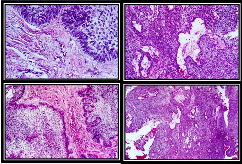
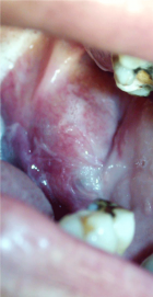
Figure 5:
Basaloid - Basal cell follicular; the microscopy was represented by keratinized squamous epithelial tissue with vacuolar degenerations, covering fibrous connective tissue that exhibits chronic inflammatory reaction, extensive areas of hemorrhage (hematoma) and mucosal salivary glands.
Read Full Article HTML DOI: 10.29328/journal.jcmei.1001003 Cite this Article Read Full Article PDF
More Images
Similar Articles
-
Cystic Micronodular Thymoma. Report of a CaseMona Mlika*,Adel Marghli ,Faouzi Mezni. Cystic Micronodular Thymoma. Report of a Case . . 2017 doi: 10.29328/journal.jcmei.1001001; 1: 001-004
-
Pulmonary Infarction Mimicking An Aspergilloma In A Heart Transplant RecipientAntonacci F*,Belliato M,Bortolotto C,Di Perna D,Dore R,Orlandoni G,D’Armini AM. Pulmonary Infarction Mimicking An Aspergilloma In A Heart Transplant Recipient . . 2017 doi: 10.29328/journal.jcmei.1001002; 1: 005-006
-
Recurrent Peripheral Ameloblastoma of the Mandible: A Case ReportAngela Jordão Camargo*,Mayara Cheade,Celso Martinelli,Plauto Christopher Aranha Watanabe. Recurrent Peripheral Ameloblastoma of the Mandible: A Case Report. . 2017 doi: 10.29328/journal.jcmei.1001003; 1: 007-010
-
Tumours of the Uterine Corpus: A Histopathological and Prognostic Evaluation Preliminary of 429 PatientsJorge F Cameselle-Teijeiro*,Javier Valdés-Pons,Lucía Cameselle-Cortizo,Isaura Fernández-Pérez,MaríaJosé Lamas-González,Sabela Iglesias-Faustino,Elena Figueiredo Alonso,María-Emilia Cortizo-Torres,María-Concepción Agras-Suárez,Araceli Iglesias-Salgado,Marta Salgado-Costas,Susana Friande-Pereira,Fernando C Schmitt. Tumours of the Uterine Corpus: A Histopathological and Prognostic Evaluation Preliminary of 429 Patients . . 2017 doi: 10.29328/journal.jcmei.1001004; 1: 011-019
-
The Risk Factors for Ankle Sprain in Cadets at a Male Military School in Iran: A Retrospective Case-control StudyFarzad Najafipour*,Farshad Najafipour,Mohammad Hassan Majlesi,Milad Darejeh. The Risk Factors for Ankle Sprain in Cadets at a Male Military School in Iran: A Retrospective Case-control Study. . 2017 doi: 10.29328/journal.jcmei.1001005; 1: 020-026
-
Magnetic Resonance Imaging Can Detect Symptomatic Patients with Facet Joint Pain. A Retrospective AnalysisWolfgang Freund*,Frank Weber,Reinhard Meier,Stephan Klessinger. Magnetic Resonance Imaging Can Detect Symptomatic Patients with Facet Joint Pain. A Retrospective Analysis. . 2017 doi: 10.29328/journal.jcmei.1001006; 1: 027-036
-
Secondary Onychomycosis Development after Cosmetic Procedure-Case ReportMariusz Dyląg*,Emilia Flisowska,Patryk Bielecki,Maria Kozioł-Gałczyńska,Weronika Jasińska. Secondary Onychomycosis Development after Cosmetic Procedure-Case Report . . 2017 doi: 10.29328/journal.jcmei.1001007; 1: 037-045
-
Andy Gump deformityPirabu Sakthivel*,Chirom Amit Singh,Chandra Sharma. Andy Gump deformity. . 2017 doi: 10.29328/journal.jcmei.1001008; 1: 046-047
-
The Death of a Baby from the Congenital Anomalies of the Urinary TractAstrit Gashi M*,Gent Sopa,Ilir Kadiri,Majlinda Bala,Petrit Pupa. The Death of a Baby from the Congenital Anomalies of the Urinary Tract. . 2018 doi: 10.29328/journal.jcmei.1001009; 2: 001-002
-
New technique of imaging cellular change to squmous cells metaplsia of cervixSalwa Samir Anter*. New technique of imaging cellular change to squmous cells metaplsia of cervix . . 2019 doi: 10.29328/journal.jcmei.1001010; 3: 001-001
Recently Viewed
-
Agriculture High-Quality Development and NutritionZhongsheng Guo*. Agriculture High-Quality Development and Nutrition. Arch Food Nutr Sci. 2024: doi: 10.29328/journal.afns.1001060; 8: 038-040
-
A Low-cost High-throughput Targeted Sequencing for the Accurate Detection of Respiratory Tract PathogenChangyan Ju, Chengbosen Zhou, Zhezhi Deng, Jingwei Gao, Weizhao Jiang, Hanbing Zeng, Haiwei Huang, Yongxiang Duan, David X Deng*. A Low-cost High-throughput Targeted Sequencing for the Accurate Detection of Respiratory Tract Pathogen. Int J Clin Virol. 2024: doi: 10.29328/journal.ijcv.1001056; 8: 001-007
-
A Comparative Study of Metoprolol and Amlodipine on Mortality, Disability and Complication in Acute StrokeJayantee Kalita*,Dhiraj Kumar,Nagendra B Gutti,Sandeep K Gupta,Anadi Mishra,Vivek Singh. A Comparative Study of Metoprolol and Amlodipine on Mortality, Disability and Complication in Acute Stroke. J Neurosci Neurol Disord. 2025: doi: 10.29328/journal.jnnd.1001108; 9: 039-045
-
Development of qualitative GC MS method for simultaneous identification of PM-CCM a modified illicit drugs preparation and its modern-day application in drug-facilitated crimesBhagat Singh*,Satish R Nailkar,Chetansen A Bhadkambekar,Suneel Prajapati,Sukhminder Kaur. Development of qualitative GC MS method for simultaneous identification of PM-CCM a modified illicit drugs preparation and its modern-day application in drug-facilitated crimes. J Forensic Sci Res. 2023: doi: 10.29328/journal.jfsr.1001043; 7: 004-010
-
A Gateway to Metal Resistance: Bacterial Response to Heavy Metal Toxicity in the Biological EnvironmentLoai Aljerf*,Nuha AlMasri. A Gateway to Metal Resistance: Bacterial Response to Heavy Metal Toxicity in the Biological Environment. Ann Adv Chem. 2018: doi: 10.29328/journal.aac.1001012; 2: 032-044
Most Viewed
-
Evaluation of Biostimulants Based on Recovered Protein Hydrolysates from Animal By-products as Plant Growth EnhancersH Pérez-Aguilar*, M Lacruz-Asaro, F Arán-Ais. Evaluation of Biostimulants Based on Recovered Protein Hydrolysates from Animal By-products as Plant Growth Enhancers. J Plant Sci Phytopathol. 2023 doi: 10.29328/journal.jpsp.1001104; 7: 042-047
-
Sinonasal Myxoma Extending into the Orbit in a 4-Year Old: A Case PresentationJulian A Purrinos*, Ramzi Younis. Sinonasal Myxoma Extending into the Orbit in a 4-Year Old: A Case Presentation. Arch Case Rep. 2024 doi: 10.29328/journal.acr.1001099; 8: 075-077
-
Feasibility study of magnetic sensing for detecting single-neuron action potentialsDenis Tonini,Kai Wu,Renata Saha,Jian-Ping Wang*. Feasibility study of magnetic sensing for detecting single-neuron action potentials. Ann Biomed Sci Eng. 2022 doi: 10.29328/journal.abse.1001018; 6: 019-029
-
Pediatric Dysgerminoma: Unveiling a Rare Ovarian TumorFaten Limaiem*, Khalil Saffar, Ahmed Halouani. Pediatric Dysgerminoma: Unveiling a Rare Ovarian Tumor. Arch Case Rep. 2024 doi: 10.29328/journal.acr.1001087; 8: 010-013
-
Physical activity can change the physiological and psychological circumstances during COVID-19 pandemic: A narrative reviewKhashayar Maroufi*. Physical activity can change the physiological and psychological circumstances during COVID-19 pandemic: A narrative review. J Sports Med Ther. 2021 doi: 10.29328/journal.jsmt.1001051; 6: 001-007

HSPI: We're glad you're here. Please click "create a new Query" if you are a new visitor to our website and need further information from us.
If you are already a member of our network and need to keep track of any developments regarding a question you have already submitted, click "take me to my Query."


