Figure 1
Occipital lobe ependymal cyst with unusual presentation
Okacha Naama*, Abdelilah Idir and Omar Boulahroud
Published: 19 September, 2019 | Volume 3 - Issue 1 | Pages: 009-011
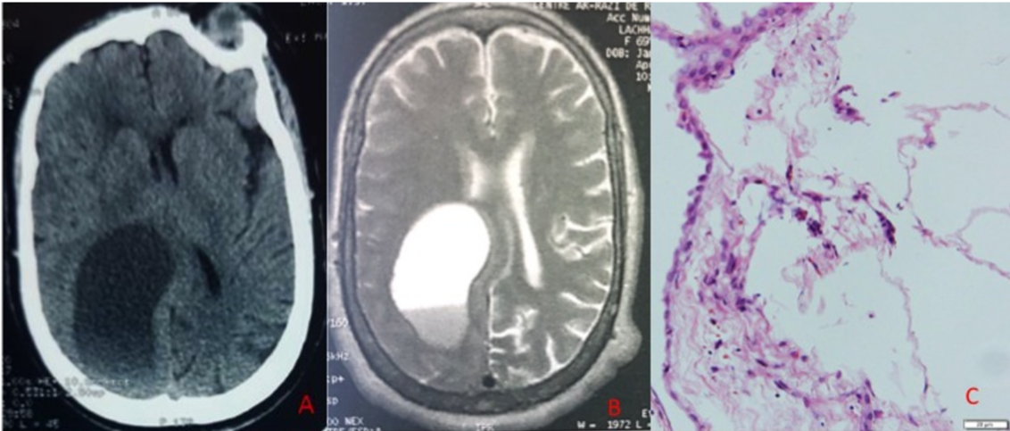
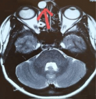
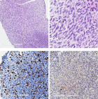
Figure 1:
(A) Contrast enhanced CT scan. A non-enhancing low density mass lesion is present in the right occipital region. (B) Initial MRI in T2 weighted axial view showing a large cystic lesion in the occipital lobe with mass effect on the lateral ventricle and adjacent structures. (C) The H&E stained slides show fragments of fibrous tissue lined by flattened to cuboidal epithelium without prominent cilia.
Read Full Article HTML DOI: 10.29328/journal.jcmei.1001012 Cite this Article Read Full Article PDF
More Images
Similar Articles
-
Cystic Micronodular Thymoma. Report of a CaseMona Mlika*,Adel Marghli ,Faouzi Mezni. Cystic Micronodular Thymoma. Report of a Case . . 2017 doi: 10.29328/journal.jcmei.1001001; 1: 001-004
-
Pulmonary Infarction Mimicking An Aspergilloma In A Heart Transplant RecipientAntonacci F*,Belliato M,Bortolotto C,Di Perna D,Dore R,Orlandoni G,D’Armini AM. Pulmonary Infarction Mimicking An Aspergilloma In A Heart Transplant Recipient . . 2017 doi: 10.29328/journal.jcmei.1001002; 1: 005-006
-
Recurrent Peripheral Ameloblastoma of the Mandible: A Case ReportAngela Jordão Camargo*,Mayara Cheade,Celso Martinelli,Plauto Christopher Aranha Watanabe. Recurrent Peripheral Ameloblastoma of the Mandible: A Case Report. . 2017 doi: 10.29328/journal.jcmei.1001003; 1: 007-010
-
Tumours of the Uterine Corpus: A Histopathological and Prognostic Evaluation Preliminary of 429 PatientsJorge F Cameselle-Teijeiro*,Javier Valdés-Pons,Lucía Cameselle-Cortizo,Isaura Fernández-Pérez,MaríaJosé Lamas-González,Sabela Iglesias-Faustino,Elena Figueiredo Alonso,María-Emilia Cortizo-Torres,María-Concepción Agras-Suárez,Araceli Iglesias-Salgado,Marta Salgado-Costas,Susana Friande-Pereira,Fernando C Schmitt. Tumours of the Uterine Corpus: A Histopathological and Prognostic Evaluation Preliminary of 429 Patients . . 2017 doi: 10.29328/journal.jcmei.1001004; 1: 011-019
-
The Risk Factors for Ankle Sprain in Cadets at a Male Military School in Iran: A Retrospective Case-control StudyFarzad Najafipour*,Farshad Najafipour,Mohammad Hassan Majlesi,Milad Darejeh. The Risk Factors for Ankle Sprain in Cadets at a Male Military School in Iran: A Retrospective Case-control Study. . 2017 doi: 10.29328/journal.jcmei.1001005; 1: 020-026
-
Magnetic Resonance Imaging Can Detect Symptomatic Patients with Facet Joint Pain. A Retrospective AnalysisWolfgang Freund*,Frank Weber,Reinhard Meier,Stephan Klessinger. Magnetic Resonance Imaging Can Detect Symptomatic Patients with Facet Joint Pain. A Retrospective Analysis. . 2017 doi: 10.29328/journal.jcmei.1001006; 1: 027-036
-
Secondary Onychomycosis Development after Cosmetic Procedure-Case ReportMariusz Dyląg*,Emilia Flisowska,Patryk Bielecki,Maria Kozioł-Gałczyńska,Weronika Jasińska. Secondary Onychomycosis Development after Cosmetic Procedure-Case Report . . 2017 doi: 10.29328/journal.jcmei.1001007; 1: 037-045
-
Andy Gump deformityPirabu Sakthivel*,Chirom Amit Singh,Chandra Sharma. Andy Gump deformity. . 2017 doi: 10.29328/journal.jcmei.1001008; 1: 046-047
-
The Death of a Baby from the Congenital Anomalies of the Urinary TractAstrit Gashi M*,Gent Sopa,Ilir Kadiri,Majlinda Bala,Petrit Pupa. The Death of a Baby from the Congenital Anomalies of the Urinary Tract. . 2018 doi: 10.29328/journal.jcmei.1001009; 2: 001-002
-
New technique of imaging cellular change to squmous cells metaplsia of cervixSalwa Samir Anter*. New technique of imaging cellular change to squmous cells metaplsia of cervix . . 2019 doi: 10.29328/journal.jcmei.1001010; 3: 001-001
Recently Viewed
-
Agriculture High-Quality Development and NutritionZhongsheng Guo*. Agriculture High-Quality Development and Nutrition. Arch Food Nutr Sci. 2024: doi: 10.29328/journal.afns.1001060; 8: 038-040
-
A Low-cost High-throughput Targeted Sequencing for the Accurate Detection of Respiratory Tract PathogenChangyan Ju, Chengbosen Zhou, Zhezhi Deng, Jingwei Gao, Weizhao Jiang, Hanbing Zeng, Haiwei Huang, Yongxiang Duan, David X Deng*. A Low-cost High-throughput Targeted Sequencing for the Accurate Detection of Respiratory Tract Pathogen. Int J Clin Virol. 2024: doi: 10.29328/journal.ijcv.1001056; 8: 001-007
-
A Comparative Study of Metoprolol and Amlodipine on Mortality, Disability and Complication in Acute StrokeJayantee Kalita*,Dhiraj Kumar,Nagendra B Gutti,Sandeep K Gupta,Anadi Mishra,Vivek Singh. A Comparative Study of Metoprolol and Amlodipine on Mortality, Disability and Complication in Acute Stroke. J Neurosci Neurol Disord. 2025: doi: 10.29328/journal.jnnd.1001108; 9: 039-045
-
Development of qualitative GC MS method for simultaneous identification of PM-CCM a modified illicit drugs preparation and its modern-day application in drug-facilitated crimesBhagat Singh*,Satish R Nailkar,Chetansen A Bhadkambekar,Suneel Prajapati,Sukhminder Kaur. Development of qualitative GC MS method for simultaneous identification of PM-CCM a modified illicit drugs preparation and its modern-day application in drug-facilitated crimes. J Forensic Sci Res. 2023: doi: 10.29328/journal.jfsr.1001043; 7: 004-010
-
A Gateway to Metal Resistance: Bacterial Response to Heavy Metal Toxicity in the Biological EnvironmentLoai Aljerf*,Nuha AlMasri. A Gateway to Metal Resistance: Bacterial Response to Heavy Metal Toxicity in the Biological Environment. Ann Adv Chem. 2018: doi: 10.29328/journal.aac.1001012; 2: 032-044
Most Viewed
-
Evaluation of Biostimulants Based on Recovered Protein Hydrolysates from Animal By-products as Plant Growth EnhancersH Pérez-Aguilar*, M Lacruz-Asaro, F Arán-Ais. Evaluation of Biostimulants Based on Recovered Protein Hydrolysates from Animal By-products as Plant Growth Enhancers. J Plant Sci Phytopathol. 2023 doi: 10.29328/journal.jpsp.1001104; 7: 042-047
-
Sinonasal Myxoma Extending into the Orbit in a 4-Year Old: A Case PresentationJulian A Purrinos*, Ramzi Younis. Sinonasal Myxoma Extending into the Orbit in a 4-Year Old: A Case Presentation. Arch Case Rep. 2024 doi: 10.29328/journal.acr.1001099; 8: 075-077
-
Feasibility study of magnetic sensing for detecting single-neuron action potentialsDenis Tonini,Kai Wu,Renata Saha,Jian-Ping Wang*. Feasibility study of magnetic sensing for detecting single-neuron action potentials. Ann Biomed Sci Eng. 2022 doi: 10.29328/journal.abse.1001018; 6: 019-029
-
Pediatric Dysgerminoma: Unveiling a Rare Ovarian TumorFaten Limaiem*, Khalil Saffar, Ahmed Halouani. Pediatric Dysgerminoma: Unveiling a Rare Ovarian Tumor. Arch Case Rep. 2024 doi: 10.29328/journal.acr.1001087; 8: 010-013
-
Physical activity can change the physiological and psychological circumstances during COVID-19 pandemic: A narrative reviewKhashayar Maroufi*. Physical activity can change the physiological and psychological circumstances during COVID-19 pandemic: A narrative review. J Sports Med Ther. 2021 doi: 10.29328/journal.jsmt.1001051; 6: 001-007

HSPI: We're glad you're here. Please click "create a new Query" if you are a new visitor to our website and need further information from us.
If you are already a member of our network and need to keep track of any developments regarding a question you have already submitted, click "take me to my Query."


