Figure 1
Aortic dissection complicating carotid dissection and myocardial infarction
Sung-Yuan Hu*, Chun-Cheng Chen and Hung-Wen Tsai
Published: 18 March, 2021 | Volume 5 - Issue 1 | Pages: 003-003

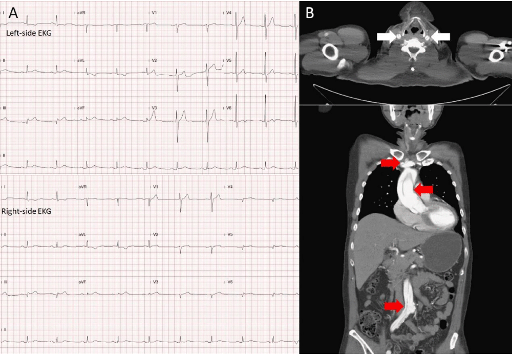
Figure 1:
Left-side electrocardiogram (EKG) revealed ST-segment elevation in II, III, and aVF, with reciprocal changes in I and aVL, disclosing inferior wall myocardial infarction. Right-side (EKG) showed ST-segment elevation in V4, disclosing right ventricular involvement (Figure 1A). The axial and coronary views of computed tomographic angiography demonstrated dissection of bilateral carotid arteries (white arrows in Figure 1B) and dissection of ascending aorta with extending to subclavian and right common iliac arteries (red arrows in Figure 1B).
Read Full Article HTML DOI: 10.29328/journal.jcmei.1001019 Cite this Article Read Full Article PDF
More Images
Similar Articles
-
Cystic Micronodular Thymoma. Report of a CaseMona Mlika*,Adel Marghli ,Faouzi Mezni. Cystic Micronodular Thymoma. Report of a Case . . 2017 doi: 10.29328/journal.jcmei.1001001; 1: 001-004
-
Pulmonary Infarction Mimicking An Aspergilloma In A Heart Transplant RecipientAntonacci F*,Belliato M,Bortolotto C,Di Perna D,Dore R,Orlandoni G,D’Armini AM. Pulmonary Infarction Mimicking An Aspergilloma In A Heart Transplant Recipient . . 2017 doi: 10.29328/journal.jcmei.1001002; 1: 005-006
-
Recurrent Peripheral Ameloblastoma of the Mandible: A Case ReportAngela Jordão Camargo*,Mayara Cheade,Celso Martinelli,Plauto Christopher Aranha Watanabe. Recurrent Peripheral Ameloblastoma of the Mandible: A Case Report. . 2017 doi: 10.29328/journal.jcmei.1001003; 1: 007-010
-
Tumours of the Uterine Corpus: A Histopathological and Prognostic Evaluation Preliminary of 429 PatientsJorge F Cameselle-Teijeiro*,Javier Valdés-Pons,Lucía Cameselle-Cortizo,Isaura Fernández-Pérez,MaríaJosé Lamas-González,Sabela Iglesias-Faustino,Elena Figueiredo Alonso,María-Emilia Cortizo-Torres,María-Concepción Agras-Suárez,Araceli Iglesias-Salgado,Marta Salgado-Costas,Susana Friande-Pereira,Fernando C Schmitt. Tumours of the Uterine Corpus: A Histopathological and Prognostic Evaluation Preliminary of 429 Patients . . 2017 doi: 10.29328/journal.jcmei.1001004; 1: 011-019
-
The Risk Factors for Ankle Sprain in Cadets at a Male Military School in Iran: A Retrospective Case-control StudyFarzad Najafipour*,Farshad Najafipour,Mohammad Hassan Majlesi,Milad Darejeh. The Risk Factors for Ankle Sprain in Cadets at a Male Military School in Iran: A Retrospective Case-control Study. . 2017 doi: 10.29328/journal.jcmei.1001005; 1: 020-026
-
Magnetic Resonance Imaging Can Detect Symptomatic Patients with Facet Joint Pain. A Retrospective AnalysisWolfgang Freund*,Frank Weber,Reinhard Meier,Stephan Klessinger. Magnetic Resonance Imaging Can Detect Symptomatic Patients with Facet Joint Pain. A Retrospective Analysis. . 2017 doi: 10.29328/journal.jcmei.1001006; 1: 027-036
-
Secondary Onychomycosis Development after Cosmetic Procedure-Case ReportMariusz Dyląg*,Emilia Flisowska,Patryk Bielecki,Maria Kozioł-Gałczyńska,Weronika Jasińska. Secondary Onychomycosis Development after Cosmetic Procedure-Case Report . . 2017 doi: 10.29328/journal.jcmei.1001007; 1: 037-045
-
Andy Gump deformityPirabu Sakthivel*,Chirom Amit Singh,Chandra Sharma. Andy Gump deformity. . 2017 doi: 10.29328/journal.jcmei.1001008; 1: 046-047
-
The Death of a Baby from the Congenital Anomalies of the Urinary TractAstrit Gashi M*,Gent Sopa,Ilir Kadiri,Majlinda Bala,Petrit Pupa. The Death of a Baby from the Congenital Anomalies of the Urinary Tract. . 2018 doi: 10.29328/journal.jcmei.1001009; 2: 001-002
-
New technique of imaging cellular change to squmous cells metaplsia of cervixSalwa Samir Anter*. New technique of imaging cellular change to squmous cells metaplsia of cervix . . 2019 doi: 10.29328/journal.jcmei.1001010; 3: 001-001
Recently Viewed
-
The value of medicine innovationRehan Haider*. The value of medicine innovation. Arch Pharm Pharma Sci. 2022: doi: 10.29328/journal.apps.1001034; 6: 030-034
-
Status of hemodialysis patients using complementary and alternative medicine practices during the COVID-19 pandemicSevil Güler*,Seda Şahan. Status of hemodialysis patients using complementary and alternative medicine practices during the COVID-19 pandemic. Arch Pharm Pharma Sci. 2022: doi: 10.29328/journal.apps.1001033; 6: 024-029
-
Cancer Cell Resistance: The Emergent Intelligence of Adaptation and the Need for Biophysical IntegrationMohamed H Doweidar*. Cancer Cell Resistance: The Emergent Intelligence of Adaptation and the Need for Biophysical Integration. Int J Clin Microbiol Biochem Technol. 2025: doi: 10.29328/journal.ijcmbt.1001031; 8: 007-008
-
Evolution of Antifungal Activity of Artemisia herba-alba Extracts on Growth of Aspergillus sp. and Rhizopus sp.Eman MG Gebreil,Nagwa SA Alraaydi,Saleh HM EL-Majberi,Idress Hamad Attitalla*. Evolution of Antifungal Activity of Artemisia herba-alba Extracts on Growth of Aspergillus sp. and Rhizopus sp.. Int J Clin Microbiol Biochem Technol. 2025: doi: 10.29328/journal.ijcmbt.1001030; 8: 001-006
-
Experience in optimizing the accessibility of services for tuberculosis in the Republic of TajikistanBobokhojaev OI**. Experience in optimizing the accessibility of services for tuberculosis in the Republic of Tajikistan. J Community Med Health Solut. 2022: doi: 10.29328/journal.jcmhs.1001022; 3: 064-068
Most Viewed
-
Feasibility study of magnetic sensing for detecting single-neuron action potentialsDenis Tonini,Kai Wu,Renata Saha,Jian-Ping Wang*. Feasibility study of magnetic sensing for detecting single-neuron action potentials. Ann Biomed Sci Eng. 2022 doi: 10.29328/journal.abse.1001018; 6: 019-029
-
Evaluation of In vitro and Ex vivo Models for Studying the Effectiveness of Vaginal Drug Systems in Controlling Microbe Infections: A Systematic ReviewMohammad Hossein Karami*, Majid Abdouss*, Mandana Karami. Evaluation of In vitro and Ex vivo Models for Studying the Effectiveness of Vaginal Drug Systems in Controlling Microbe Infections: A Systematic Review. Clin J Obstet Gynecol. 2023 doi: 10.29328/journal.cjog.1001151; 6: 201-215
-
Prospective Coronavirus Liver Effects: Available KnowledgeAvishek Mandal*. Prospective Coronavirus Liver Effects: Available Knowledge. Ann Clin Gastroenterol Hepatol. 2023 doi: 10.29328/journal.acgh.1001039; 7: 001-010
-
Causal Link between Human Blood Metabolites and Asthma: An Investigation Using Mendelian RandomizationYong-Qing Zhu, Xiao-Yan Meng, Jing-Hua Yang*. Causal Link between Human Blood Metabolites and Asthma: An Investigation Using Mendelian Randomization. Arch Asthma Allergy Immunol. 2023 doi: 10.29328/journal.aaai.1001032; 7: 012-022
-
An algorithm to safely manage oral food challenge in an office-based setting for children with multiple food allergiesNathalie Cottel,Aïcha Dieme,Véronique Orcel,Yannick Chantran,Mélisande Bourgoin-Heck,Jocelyne Just. An algorithm to safely manage oral food challenge in an office-based setting for children with multiple food allergies. Arch Asthma Allergy Immunol. 2021 doi: 10.29328/journal.aaai.1001027; 5: 030-037

HSPI: We're glad you're here. Please click "create a new Query" if you are a new visitor to our website and need further information from us.
If you are already a member of our network and need to keep track of any developments regarding a question you have already submitted, click "take me to my Query."


